3D printing of Bioactive Implants: Engineering the Interplay of Materials and Design
Published 24 September, 2025
In a new study published in Biomedical Technology, a new 3D printing approach that could transform the way surgeons repair damaged bone was reported. The study shows how adjusting both the printing ink and the way material is laid down can change the strength and healing potential of implants. The researchers used a technique called direct ink writing, which works at room temperature to print dense, solid implants. Unlike many existing 3D-printed scaffolds that are porous and fragile, these implants are mechanically stable while still encouraging bone cells to grow and form new tissue.
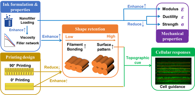
A key finding was that implants printed at different angles behaved in surprising ways. “In fused deposition modelling, a common 3D printing method, printing filaments in the same direction as the applied force usually makes the implant stronger,” explains lead author Hongyi Chen from University College London. “But with our approach, we found the opposite—implants printed at 90 degrees actually had better strength because the filaments bonded more effectively.”
Further, the researchers added tiny particles of Laponite, a nanoclay, into the printing ink. “These particles made the ink thicker, helping the printed shapes hold their form, while also releasing bioactive ions that encourage bone cells to attach and grow,” says Chen.
Tests showed that implants with higher Laponite content had a 110% increase in stiffness compared to pure polymer implants, and bone-forming cells on these implants showed greater proliferation and mineralization over time.
“What makes this study distinctive is that we didn’t just look at one factor in isolation,” adds Chen. “By examining the interplay between ink composition, printing orientation, structure, mechanical behaviour, and cell response, we could see how design choices at each stage influence the final biological outcome.”
The work highlights a new strategy for creating bone implants that balance mechanical stability with bioactivity, offering potential benefits for areas such as craniomaxillofacial reconstruction and dental bone grafting. The researchers revealed that the next steps will involve exploring porous and more complex designs, as well as testing in preclinical models. If successful, the approach could enable patient-specific implants produced quickly in hospital labs or even at the point of care.

Contact author:
Hongyi Chen, Post-doctoral research fellow at University College London, hongyi.chen.16@ucl.ac.uk
Funder:
Support from the Engineering and Physical Sciences Research Council (EPSRC) Manufacturing the Future programme (no. EP/N024915/1) to SB for part of this work is gratefully acknowledged.
Conflict of interest:
The authors declare no conflict of interest
See the article:
Chen, H.*, Cheng, R., Chung, S. H., Marghoub, A., Zhong, H., Fang, G., Balabani, S., Di-Silvio, L., & Huang, J.* (2025). Direct ink writing of bioactive PCL/laponite bone Implants: Engineering the interplay of design, process, structure, and function. Biomedical Technology, 11, 100101.
https://doi.org/10.1016/j.bmt.2025.100101

